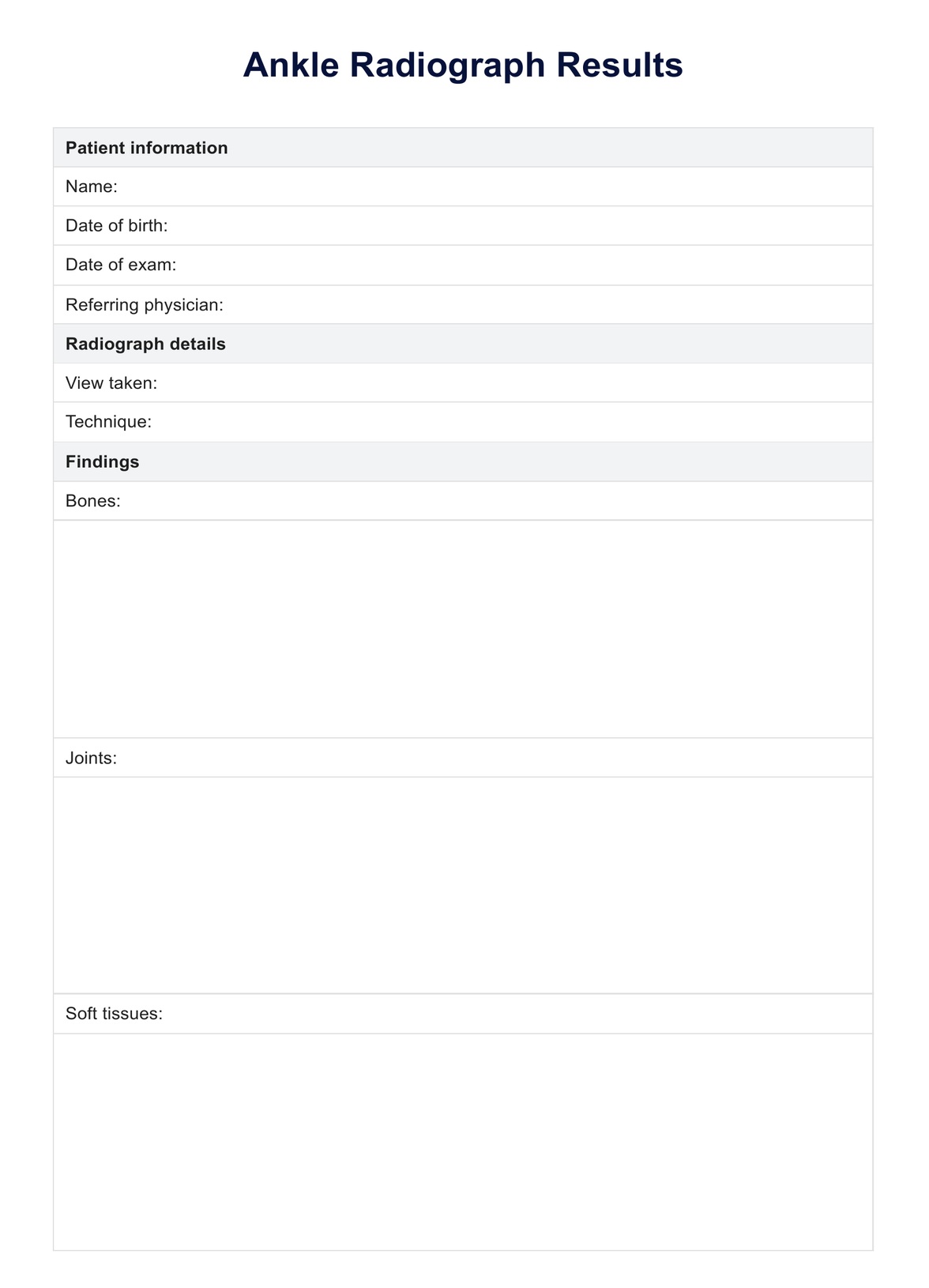
What is an ankle radiograph?
An ankle radiograph, commonly known as an ankle X-ray, is a diagnostic imaging technique used to visualize the bones and joints of the ankle. This test helps medical professionals assess the ankle for fractures, dislocations, arthritis, and other conditions. The radiograph uses a small amount of ionizing radiation to produce images of the ankle, which can show the alignment of the bones and the condition of the joint spaces and surrounding tissues.
Purpose
The primary purpose of an ankle radiograph is to evaluate the ankle following injury to diagnose fractures or dislocations. It is also used to monitor the progression of diseases such as osteoarthritis or rheumatoid arthritis.
Additionally, ankle radiographs can help plan medical or surgical treatments and assess healing after a fracture or surgery. The radiographs evaluate the condition of both the lateral and medial malleoli, helping diagnose fractures and track disease progression.
The three views of ankle radiographs
Ankle radiographs typically consist of three views, which provide different angles for thorough examination:
- Anteroposterior (AP): This is the frontal view of the ankle. The X-ray beam passes from the front to the back of the ankle. It helps assess the alignment of the tibia, fibula, talus, and joint spaces. The AP view is especially valuable for assessing the alignment of the distal tibia with the talus and the tibial articular surface.
- Mortise: This view is similar to the AP view, but the foot is rotated slightly. This view is particularly important for evaluating the ankle mortise, a U-shaped notch between the tibia and the fibula where the talus fits. It provides a clear image of the joint space and the integrity of the ankle mortise. The mortise view also provides a detailed examination of the ankle mortise, focusing on the alignment and space between the lateral malleolus and medial malleolus.
- Lateral: The X-ray beam passes from one side of the ankle to the other. This side view helps visualize the ankle's overlapping bones, the alignment of the tibia and fibula with the talus, and the posterior and anterior aspects of the ankle joint. The lateral view not only helps visualize the distal fibula but also provides insight into the condition of the proximal fibula.
These three views are crucial for a comprehensive assessment of the ankle, allowing for accurate diagnosis and appropriate management of ankle-related conditions.
Preparations for ankle radiographs
Several important steps must be taken when preparing for an ankle radiograph to ensure a smooth imaging process and accurate results. First, patients are asked to remove any clothing that covers the ankle area, along with all jewelry and metal objects that might obscure the ankle's view or interfere with the X-ray imaging. Sometimes, patients may be given a hospital gown to wear during the procedure.
Positioning for the X-ray is critical for capturing the best possible image. The radiologic technologist will guide the patient on positioning their ankle for each required view. For the anteroposterior view, the foot is placed flat and facing forward. The foot is slightly rotated inward for the mortise view, and for the lateral view, the patient is asked to turn the foot sideways.
Additionally, protective measures are often taken to minimize radiation exposure. Protective lead aprons or shields are used to cover areas of the body not being imaged, such as the pelvis, particularly in pregnant or reproductive-age patients.
It is also crucial for the patient to remain still during the X-ray to prevent the images from blurring. The technologist will help the patient find a comfortable yet firm position that can be maintained for a few seconds while the X-ray is taken. These steps are essential for ensuring that the ankle radiograph is clear and detailed, providing valuable assistance in accurate diagnosis and treatment planning.
How do professionals convey ankle radiograph results?
Once an ankle radiograph is taken, a radiologist specializing in interpreting medical images will review the X-rays. If available, they will look for any signs of injury, disease, or abnormality in the ankle and compare them with previous images. Part of the assessment includes examining the talar dome for smoothness and integrity, which is crucial for ankle mobility and stability.
The radiologist will then write a detailed report outlining their findings, noting any changes and potential concerns. The radiologist also assesses for any signs of ligamentous injury, which are crucial for determining the full extent of ankle trauma. They also look at the integrity of the deltoid ligament, particularly its lateral aspect, for any signs of injury or strain.
The report is sent to the physician who requested the X-ray. Depending on the context of the examination, this might be the patient’s general practitioner, an emergency room physician, or an orthopedic specialist. The referring physician will review the radiologist's report and may consult with the radiologist for further clarification or discussion about the findings.
The final step involves the physician discussing the results with the patient. During this consultation, the physician explains the radiograph findings, what they mean for the patient's health, and the next steps in terms of treatment or further testing. This discussion is crucial as it helps the patient understand their medical condition and the implications of the radiograph results, ensuring they are fully informed about their health and care options.


