How to test for tarsal navicular stress fractures
Testing for tarsal navicular stress fractures involves a combination of clinical evaluation and imaging studies to diagnose the condition accurately. Here's a step-by-step guide on how healthcare providers typically test for this type of tarsal navicular stress fracture progression of an injury or fracture:
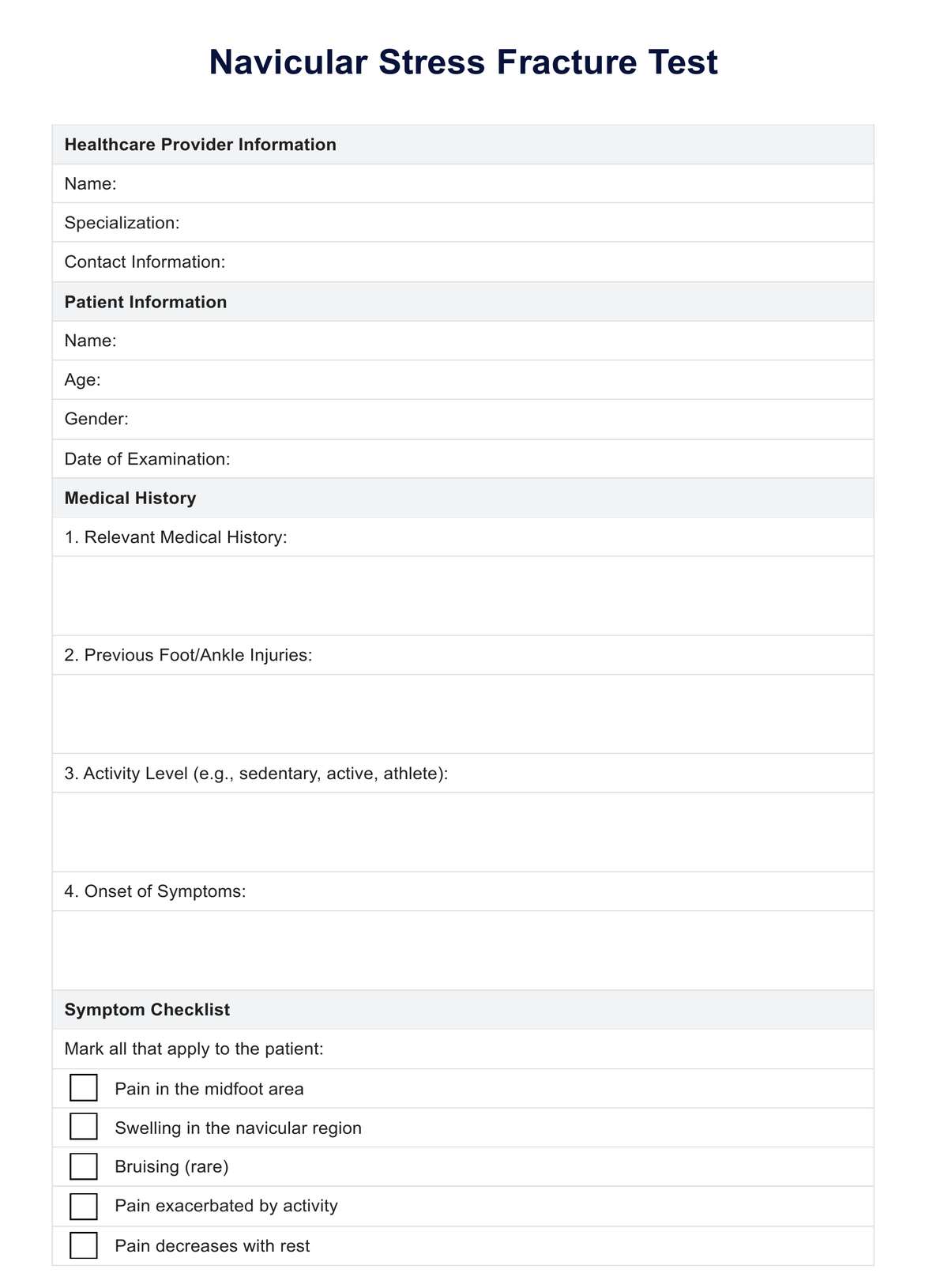
Clinical evaluation
A thorough evaluation is needed to test for navicular stress fracture:
- Patient history: The healthcare provider will start by gathering a detailed history, focusing on any recent increases in activity level, changes in training routines, or specific incidents that could have led to injury.
- Physical examination: The examination will focus on the midfoot area, especially over the navicular bone. The provider will look for tenderness, swelling, or bruising. They may also perform specific maneuvers, such as the "navicular squeeze" or "hop test," to reproduce the symptoms and localize the pain.
Imaging studies
After a clinical evaluation, imaging tests and bone scans are crucial to confirm the diagnosis, as navicular stress fractures can be difficult to detect with physical examination alone.
- X-rays: Initially, X-rays may be taken to rule out other causes of foot pain. However, navicular stress fractures often do not appear on X-rays until the healing process has begun, which might be weeks after the onset of symptoms.
- Magnetic Resonance Imaging (MRI): MRI is more sensitive and can detect navicular stress fractures earlier than X-rays. It can show both the bone and soft tissue, providing a detailed image that helps confirm the diagnosis.
- Computed tomography (CT) scan: A CT scan may be used to assess the fracture's extent and provide detailed images of the bone structure. It's particularly useful in planning treatment for complex fractures or involving significant bone displacement.
- Bone scan: Though less commonly used now due to the availability of MRI, a bone scan might be considered if MRI is not available. It involves injecting a small amount of radioactive material into the bloodstream, accumulating in areas of increased bone activity, such as a fracture site.
Follow-up and monitoring
After initial diagnosis, patients with a tarsal navicular stress fracture will typically undergo a period of rest and immobilization, with follow-up imaging to monitor the healing process. The choice of imaging for follow-up will depend on the initial findings and the healthcare provider's clinical judgment.
The correct diagnosis and treatment plan for a tarsal navicular stress fracture are crucial for optimal recovery from a complete fracture and for the prevention of complications such as nonunion or chronic pain.
You can use the Clinical Evaluation Template and Workout Planner Template to bolster your practice and optimize client results. Implementing both tools ensures a thorough approach to both health assessments and exercise planning.