Shingles Nerve Pathways Map
Looking for a Shingles Nerve Pathways Map? Get the gist of shingles and its pathways by downloading our handout.
Use Template
## **Shingles, nerve paths, and dermatomes**
Shingles, caused by the Varicella zoster virus, is a reactivation of the same virus that causes chickenpox. This herpes zoster infection affects nerve pathways and can lead to significant complications, such as nerve pain and postherpetic neuralgia.
The spinal cord and spinal nerve pathways play a significant role in how the herpes zoster shingles virus spreads. Once the Varicella zoster virus reactivates, it travels along the sensory nerves, often affecting dermatomes, which are specific areas of skin supplied by a single spinal nerve root (Maher N & Mounir N, 2023). The shingles rash typically follows these dermatomes, appearing as a painful blistering skin eruption.
Cranial nerves, particularly the trigeminal nerve, can also be affected, leading to conditions like herpes zoster ophthalmicus, where the virus impacts the eye area (Bunya, 2024).
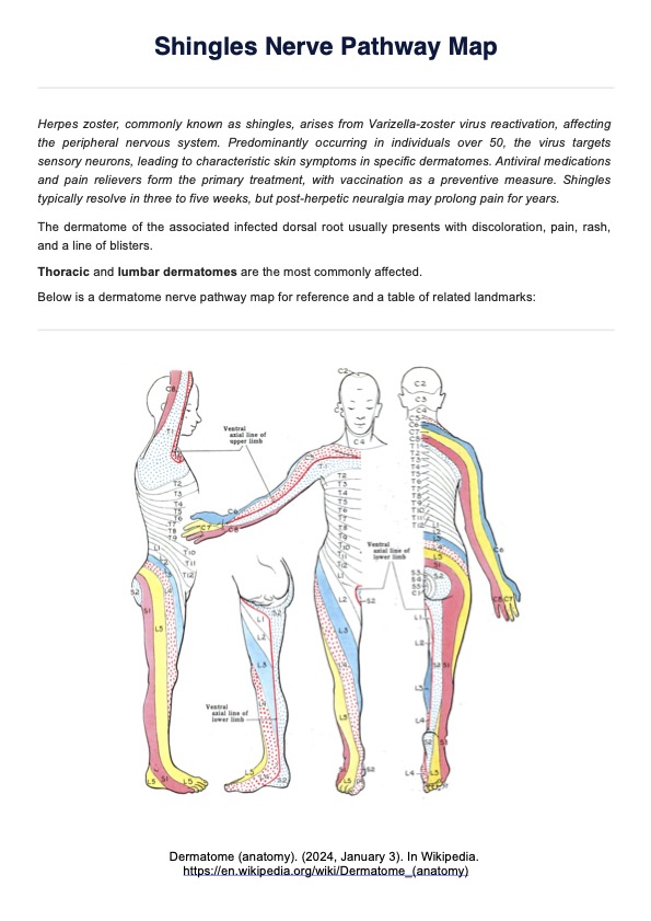
Recognizing the distinct patterns in the Shingles Nerve Pathways Map helps healthcare providers anticipate and manage complications associated with the virus effectively. Understanding these connections can improve patient outcomes and enhance the management of this painful condition.
### **How do shingles affect dermatomes?**
Shingles, also called herpes zoster, results from the reactivation of the Varicella zoster virus within the spinal cord or cranial nerve pathways. This viral infection typically follows spinal nerve distributions known as dermatomes, which are specific skin areas linked to a single spinal nerve. When the herpes zoster virus reactivates, it travels along the affected spinal nerves, causing a characteristic shingles rash in the associated dermatome.
The burning pain and severe pain often felt in the area precede the rash and are due to the nerve's irritation. In cases where the cranial nerve is involved, like the facial nerve, the condition is referred to as herpes zoster ophthalmicus, which may impact the eye and surrounding structures (Minor et al., 2025). Healthcare providers must recognize this herpes zoster shingles pattern to diagnose shingles — alongside a [Shingles Test](https://www.carepatron.com/templates/shingles/) — and manage potential complications such as post-herpetic neuralgia, a persistent pain that occurs after the rash resolves.
Shingles Nerve Pathways Map Template
## **What is a Shingle Nerve Pathways Map?**
A Shingles Nerve Pathways Map depicts how the Varicella zoster virus affects the peripheral nervous system, causing a painful rash along specific nerve pathways. When shingles (herpes zoster) reactivates, the virus spreads along sensory nerve fibers. These fibers are part of the peripheral nervous system and transmit sensory information, including shingles pain, to the brain.
### **Common Shingles Nerve Pathways Map**
A Shingles Nerve Pathways Map typically follows the dermatomes, which are skin areas innervated by individual spinal or cranial nerves. The most frequently affected nerve pathways in herpes zoster involve the thoracic, cervical, and trigeminal nerves.
- **Thoracic nerve pathway**: The thoracic dermatomes (T1-T12) are the most commonly affected in shingles. The spinal nerves exit the thoracic spine and wrap around the torso, leading to a painful rash along the chest and upper back. This presentation resembles a band or stripe around the body, often mistaken for a heart or lung issue due to the location.
- **Cervical nerve pathway**: In some cases, the cervical nerves (C2-C8) are affected, leading to shingles outbreaks along the neck, upper shoulders, and arms. The involvement of these sensory nerve fibers can cause radiating pain and discomfort in the neck and arm areas.
- **Trigeminal nerve pathway**: When the cranial nerve (particularly the trigeminal nerve) is involved, it can result in herpes zoster ophthalmicus, affecting the eye, forehead, and scalp. This pattern is serious, as it can lead to complications like vision loss if not treated promptly.
### **Benefits of using a dermatome map for shingles**
Using a dermatome map for shingles provides numerous clinical benefits in diagnosing and treating complications arising from the reactivation of the chickenpox virus, also known as herpes zoster. A dermatome map allows healthcare practitioners to pinpoint the exact areas of the skin affected by the virus, which directly correspond to the brain and spinal cord pathways. Here are its key benefits:
- **Precise identification of affected nerves**: A [Dermatome Map](https://www.carepatron.com/templates/dermatome-map/) helps clinicians accurately identify which spinal or cranial nerves are impacted by the virus. This is crucial for targeting treatment and managing nerve pain effectively.
- **Guided treatment of nerve pain**: By correlating the shingles rash with specific dermatomes, healthcare professionals can implement more effective strategies for treating nerve pain, reducing the risk of long-term complications such as segmental zoster paresis or postherpetic neuralgia.
- **Differentiation from other viral infections**: The map helps distinguish shingles from other conditions like herpes simplex, which may present with similar symptoms but affect different nerve pathways.
- **Informed vaccination strategies**: The map supports decisions related to the zoster vaccine, allowing practitioners to target high-risk patients, especially those with weakened immune system functions.
## **How to use our Shingles Nerve Pathways Map?**
To accurately diagnose and manage herpes zoster cases, using the Shingles Nerve Pathways Map from Carepatron is essential for visualizing affected dermatomes. Follow these steps to effectively utilize the map in clinical settings.
### **Step 1: Access the Carepatron template**
Obtain the Shingles Nerve Pathways Map template from Carepatron's platform, which features the Keegan and Garret map, which you may be familiar with as it is one of the most common in textbooks -- despite there being no standardization set (Downs & Laporte, 2011). To add, it is also commonly used in clinical practice (Hoagland, 2025).
### **Step 2: Identify the affected nerve pathway**
Examine the patient's painful rash and use the map to correlate the rash distribution with the corresponding sensory nerve fibers or cranial nerve.
### **Step 3: Locate the spinal nerve exit**
Match the rash location with the dermatome on the map to determine where the spinal nerves exit the spine or which peripheral nervous system nerves are involved.
### **Step 4: Assess severity and spread**
If the rash covers multiple dermatomes, consider disseminated zoster. Use the map to check for involvement of multiple nerves or signs of central nervous system impact.
## **References**
Bunya, V. Y. (2024, July 2). Herpes Zoster Ophthalmicus. MSD Manual Professional Edition; MSD Manuals. https://www.msdmanuals.com/professional/eye-disorders/corneal-disorders/herpes-zoster-ophthalmicus
Downs, M. B., & Laporte, C. (2011). Conflicting Dermatome Maps: Educational and Clinical Implications. Journal of Orthopaedic and Sports Physical Therapy, 41(6), 427–434. https://doi.org/10.2519/jospt.2011.3506
Hoagland, T. M. (2025, February 19). Dermatomes Anatomy: Overview, Gross Anatomy, Natural Variants. Medscape.com; Medscape. https://emedicine.medscape.com/article/1878388-overview?form=fpf#a2
Maher N, G., & Mounir N, G. (2023). The Neuroanatomical Basis of Unfamiliar Presentations of Herpes Zoster: A Review and A Case Report. Clinical Medical Reviews and Case Reports, 10(3). https://doi.org/10.23937/2378-3656/1410421
Minor, M., Bharat Gurnani, & Payne, E. (2025, September 14). Herpes Zoster Ophthalmicus. Nih.gov; StatPearls Publishing. https://www.ncbi.nlm.nih.gov/books/NBK557779/
Commonly asked questions
The classic pattern runs along a nerve root from the spine to the rib and all the way to the front of the chest.
In rare cases, it may.
The map correlates the distribution of the shingles rash with specific spinal nerves, allowing clinicians to accurately diagnose affected areas and manage nerve pain effectively.


EHR and practice management software
Get started for free
*No credit card required
Free
$0/usd
Unlimited clients
Telehealth
1GB of storage
Client portal text
Automated billing and online payments