Subscapularis Anatomy DiagramAI Template IconToolbar IconShare Icon

Subscapularis Anatomy Diagram

What is the subscapularis muscle? How does it function as part of the rotator cuff muscles? Download our free anatomy diagram to gain a detailed understanding of this crucial muscle.

Olivia Sayson avatar

By Olivia Sayson on Aug 2, 2025.

Fact Checked by Ericka Pingol.

Use Template

What is the subscapularis muscle?

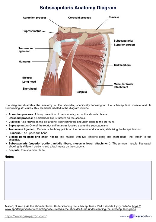
The subscapularis is one of the four rotator cuff muscles, alongside the supraspinatus, infraspinatus, and teres minor muscles. The subscapularis muscle originates from the subscapular fossa on the anterior surface of the scapula and is a large triangular muscle, the largest branch of the axillary artery, in fact.

The subscapularis tendon inserts onto the lesser tubercle of the humerus. Its primary function is the internal rotation of the arm at the shoulder joint.

The upper and lower subscapular nerves, which are branches of the posterior cord of the brachial plexus provide subscapularis muscle innervation. The subscapularis muscle is crucial for upper limb movement and stability, and injuries such as subscapularis tendon tears or a subscapularis tear can significantly impact shoulder function and may require attention similar to other rotator cuff muscles.

Subscapularis Anatomy Diagram Template

Download PDF Template

How does our Subscapularis Anatomy Diagram work?

Use the Subscapularis Anatomy Diagram template to enhance your practice, patient education, and professional collaboration. Here's how:

Step 1: Download the template

Open the Subscapularis Anatomy Diagram template by clicking "Use template," on this page, which opens it on the Carepatron app's template editor. This editor lets you customize the template before printing or filling it out. You can also get a ready-to-use PDF by clicking "Download."

Step 2: Review the diagram

Open the PDF and familiarize yourself with the detailed illustration of the subscapularis muscle, including its origin, insertion, and relationship with other rotator cuff muscles like the supraspinatus, and shoulder structures such as the clavicle, acromion process, and humerus.

Step 3: Utilize for clinical practice

Use the diagram during patient consultations to explain the anatomy of the shoulder and the role of the subscapularis muscle. This can help in discussing conditions such as subscapularis tendon tears or chronic shoulder pain.

Step 4: Educate patients and colleagues

Share the diagram with patients to help them visualize and understand their condition, the nature of their injury, or the rationale behind their treatment plan.

Step 5: Incorporate into reports and presentations

Include the diagram in your clinical reports, presentations, or educational materials to provide a clear visual reference.

Benefits of having a Subscapularis Anatomy Diagram

There are four main benefits of having a subscapularis muscle anatomy diagram:

Enhanced anatomical understanding

One of the main benefits of having a diagram is gaining a better understanding of the muscle, its precise localization, its relationship to surrounding structures, and the nerve and blood supply within the muscle.

Improved diagnosis

When a patient presents with shoulder pain, a diagram can help healthcare professionals visualize potential injury sites within the subscapularis, understand referred pain patterns, interpret imaging, and decide which tests or assessments to conduct.

Effective treatment planning

Healthcare providers can use the anatomy diagram for patient education and planning for treatments such as injections, surgery, and even rehabilitation exercises.

Enhanced communication

With an anatomy diagram, healthcare professionals have a common anatomical diagram during cases, which ensures clear and consistent communication. Aside from that, educators and students can use it for learning and teaching.

Commonly asked questions

The main action of subscapularis is internal rotation of the humerus. It also contributes to arm adduction and anterior stability of the glenohumeral joint.

Special physical exam tests like the lift-off test, bear hug test, and belly press test can detect subscapularis tears. MRI provides the definitive imaging diagnosis.

The subscapular artery, a branch of the axillary artery, is the main blood supply to the subscapularis muscle.

EHR and practice management software

Get started for free

*No credit card required

Free

$0/usd

Unlimited clients

Telehealth

1GB of storage

Client portal text

Automated billing and online payments